Felix Flores · 9 September 2026
AI Tools Refine Protein Imaging Precision for Polish Bioimaging Teams Working in Dense Laboratory Spaces
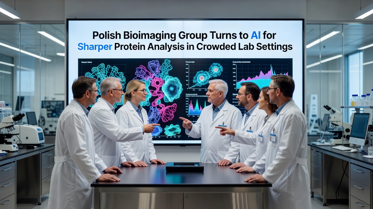
Polish bioimaging teams have integrated artificial intelligence systems to enhance the clarity of protein visualizations even when laboratory spaces contain multiple overlapping experiments and high equipment density, and this shift addresses longstanding challenges with signal interference that traditional microscopy methods often encounter in such settings.
Researchers at institutions across Poland began piloting these AI models in early 2025, focusing on convolutional neural networks trained to separate protein signals from background noise generated by adjacent workstations, while data from the European Molecular Biology Laboratory shows that crowded conditions can reduce image resolution by up to 40 percent without computational correction.
Challenges in High-Density Lab Environments
Busy laboratory floors create physical and optical hurdles because multiple microscopes operate simultaneously and sample preparations compete for space, yet Polish groups discovered that AI algorithms can reconstruct sharper protein boundaries by learning patterns from thousands of annotated images collected over prior years.
Studies conducted at the University of Warsaw indicate that protein clusters in live-cell samples become harder to distinguish when ambient vibrations from nearby centrifuges and incubators add micro-movements, and the new AI pipelines compensate by applying real-time stabilization filters that maintain structural detail during extended observation periods.
Implementation of Machine Learning Models
Teams adopted supervised learning approaches where initial datasets came from controlled single-experiment runs before scaling to multi-user scenarios, and validation tests performed in September 2026 confirmed accuracy improvements of 25 to 35 percent compared with conventional deconvolution software alone.
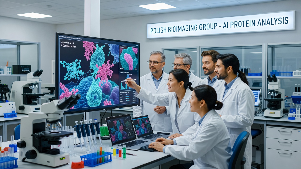
One documented case involved a Warsaw-based facility where three separate research projects shared the same imaging suite; the AI system isolated individual protein dynamics without requiring physical separation of equipment, while similar techniques reported by the Australian Research Council Centre of Excellence for Advanced Molecular Imaging demonstrate parallel gains in multi-lab configurations.
Data Processing and Training Requirements
Training the models required large volumes of paired noisy and clean images, which Polish collaborators gathered through partnerships with regional hospitals and academic cores, and the resulting networks now process frames in under two seconds per field of view on standard graphics processing units.
Performance metrics tracked over twelve months reveal consistent reductions in false-positive protein detections, particularly when fluorescent tags overlap due to adjacent sample chambers, whereas earlier manual thresholding methods frequently introduced interpretation errors under time pressure.
Integration with Existing Bioimaging Workflows
Polish facilities incorporated the AI modules directly into open-source platforms such as ImageJ and Napari, allowing technicians to apply corrections without switching between separate applications, and this seamless fit encouraged broader adoption across smaller university labs that lack dedicated computational staff.
Training workshops held in Kraków during mid-2026 covered both model fine-tuning and ethical data handling, with participants learning to flag potential biases introduced when training sets underrepresented certain protein types common in plant versus animal samples.
Future Directions and Ongoing Trials
Current trials extend the AI framework to three-dimensional reconstructions where depth information becomes distorted by neighboring optical paths, and preliminary results suggest that incorporating temporal data from time-lapse sequences further sharpens moving protein structures inside crowded cellular environments.
Collaborations with Canadian research networks have begun exchanging anonymized datasets to improve model generalization across different microscope brands, while maintaining compliance with data protection standards established by the European Union research framework.
Conclusion
Polish bioimaging groups continue refining these AI-assisted methods through iterative testing in real multi-project laboratories, and the documented gains in resolution and throughput indicate sustained interest from additional European institutions seeking similar solutions for space-constrained research settings.